

Did you know that Glasgow was home to the world’s first medical x-ray department? In March 1896, just a few months after the discovery of x-rays by the German physicist Wilhelm Röntgen, Dr John Macintyre established an x-ray unit in the electrical department of the Glasgow Royal Infirmary. You can find out more about Macintyre’s pioneering radiology work in an earlier blog post from 2014, Glasgow’s Contribution to Radiology.
Some of the early x-ray tubes from Macintyre’s radiology department are currently featured in our exhibition, Our Science and Art: Visualising the Human Body. The exhibition examines the ways in which technologies for depicting and looking inside the body have evolved and influenced medical practice from the 16th to 21st centuries. The discovery of x-rays allowed physicians and surgeons to visualise the body in an entirely new way.
There are eight tubes in total in our collection. They come in a variety of shapes, sizes and constructions, and each had a different use in the radiology department at the Royal. We’ve recently updated our catalogue records to provide a bit more information on these differences. Follow the links below to find out more:
- 1. Jackson Focus X-ray tube (c. 1895)
- 2. Early x-ray tube, similar to those used in Röntgen’s experiments (c. 1896)
- 3. Heavy current x-ray tube (c. 1918)
- 4. Another heavy current x-ray tube (c. 1920)
- 5. Geissler tube (c. 1896) – not an x-ray tube at all, but a gas discharge tube, the invention of which predates x-rays by several decades. This technology is still used today in modern neon lighting.
- 6. Small current x-ray tube (c. 1909)
- 7. Heavy current x-ray tube (c. 1918)
- 8. Valve tube (c. 1896) – another one that’s not actually an x-ray tube. These tubes were put in circuit with x-ray tubes of the gas type to suppress the harmful inverse current which was liable to occur with induction coils.
The tubes were donated to the College in 1955 by Mrs Read, the widow of Dr Charles Read, a former Fellow of the Royal Faculty who was Senior Assistant Radiologist at the Royal Infirmary until he decided to focus exclusively on dental radiology. It can be quite challenging to display objects like this, and they’ve only rarely been available to view until now. The glass is extremely fragile, but because their shape is so round they’re also prone to rolling about. We’ve had special mounts made from acrylic and foam cushioning to make sure they stay in place in our display cases! It can also be difficult for visitors to imagine how these items, now removed from their original context, actually worked. Putting more detailed information in our catalogue can help with this, but we’ve also created a short video, showing a simplified version of the heavy current tubes shown above:
And here you can (just about) see one of the bulbs in situ at the Royal Infirmary in 1914:
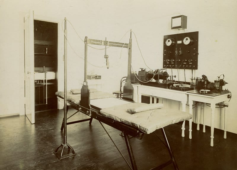
Electrical department at Glasgow Royal Infirmary c.1914
The tubes will be on display as part of Our Science and Art: Visualising the Human Body until July 2019. You can visit us on Monday afternoons between 2pm and 5pm, or at other times by appointment. We’ll also have extended opening on Monday 17 December. Check out our website for more details or to contact us.
Leave a Reply
You must be logged in to post a comment.